

Equipment
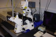
Leica LMD7000 Laser Microdissection System
To understand biological processes it is becoming increasingly important to analyse biomolecules at the resolution of single cells or cell-types. Laser Microdissection (LMD) allows you to be that “specific”.
LMD makes it possible to distinguish between relevant and non-relevant cells or tissues and enables researchers to obtain homogeneous, ultrapure samples from heterogeneous starting material. It allows you to selectively and routinely analyze regions of interest down to single cells (or even subcellular structures) to extract DNA, RNA, protein or metabolites either from living cells (cell cultures) or from fixed or frozen tissue sections in any biological system.
The Leica LMD7000 ensures contact free dissection of cells using an UV laser and collects dissected cells by gravity. High energy focussed laser pulses induce “cold ablation” of the tissue without impairing or heating the surrounding tissue. The LMD7000 is an automated upright microscope, has a high power laser with additional options to adjust the laser pulse frequency and laser head current for dissection of hard tissues like bone, wood or plant tissue. The system allows laser microdissection to be combined with standard multichannel fluorescence microscopy to aid the selection of specific cell types via histological staining, immunochemistry or use of fluorescently tagged proteins. It offers an extremely precise, highly selective laser microdissection method for a broad range of tissues, including live cell applications such as isolation of specific cells from cell cultures, cell ablation or microsurgery.
Technical details
The LMD7000 uses a 349 nm solid state diode pumped UV laser (120 µJ, pulse frequency 10 – 5000 Hz, pulse lenght < 4 ns), with continuously adjustable laser aperture, speed and focus. The laser is moved via optics.
Fully automated high-end upright microscope (DM6000B). Dedicated LMD objectives available: 5x, 10x, 20x, 40x and 63x.
Fully integrated fluorescence, allows live cutting within bright-field and fluorescence (BGR filters for DAPI, FITC, TRITC/PI) mode.
Real-time cutting can be done directly on the sample with a Pen-screen.
The use of special membrane coated glass or frame slides is recommended for mounting tissue sections, see brochure.
Samples can be collected via gravity in:
- Standard PCR tube caps 0.2 ml
- Standard PCR tube caps 0.5 ml
- 8-strip wells
- Petri dishes with or without membrane (or Leica ibidi slides)
Applications
Applications include: single cell isolation for extraction of DNA, RNA, protein or metabolites from nearly any biological system, allowing downstream (cell-specific) analyses such as for example PCR, sequencing, qRT-PCR, microarray, genetic fingerprinting, 2-D protein gels, mass-spec analyses (LC-MS/MS / MALDI). Additionally, the system can be used for isolation of specific living cells from cell cultures and for cell-ablation or microsurgery in living tissue. The system can further be used for microengraving of slides or samples for electron microscopy.
More information
- For more information about the instrument, visit the Leica website
- For more information on potential applications in life science research, visit the Leica Science lab site
In addition to the spinning disk confocal microscope, Wageningen University & Research, Shared Research Facilities offers the use of:
-
Zeiss LSM 510-META Confocal laser scanning microscope
A traditional point scanning confocal laser scanning microscope (CLSM) with excitation from 405 to 633 and multichannel imaging. The system provides... -

Spinning disk confocal microscopes
The Yokogawa Spinning Disk microscopes allow multicolor fluorescence image acquisition at high speed with low light, by using sensitive...