

facility_label
Wageningen Electron Microscopy Centre
The Wageningen Electron Microscopy Centre provides microscopy and specimen preparation facilities for biological and materials science research. In addition, the centre offers necessary training and access to all microscopes to faculty, staff, and students of Wageningen University & Research as well as external users.
Electron Microscopy introduction
Electron microscopy uses an electron beam to create an image of a sample. Because of this an electron microscope has a much greater resolving power than a light microscope and is capable of much highermagnifications (up to 2 million times).

Transmission Electron Microscopy
In transmission electron microscopy (TEM) a beam of electrons is transmitted through a thin specimen to form an image. After magnification the image is projected onto a fluorescent screen or a digital camera.

Scanning Electron Microscopy
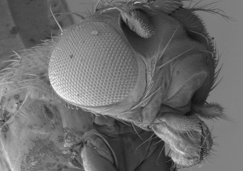
In a scanning electron microscope (SEM) an electron beam is scanning in a raster pattern over the sample. The electrons interact with atoms in the sample and the secondary electrons emitted by the sample are used to form an image of the sample surface. The SEM at WEMC is a FEI Magellan 400, with an electron energy range from 1 kV to 30 kV and a sub-nanometer resolution (0.8 nm at 15 kV—0.9 nm at 1 kV).
Electron microscopes
Electron microscopy uses an electron beam to create an image of a sample. Because of this an electron microscope has a much greater resolving power than a light microscope and is capable of much highermagnifications (up to 2 million times). There are two types of electron microscopes - transmission electron microscopes (TEM) and scanning electron microscopes (SEM).
Sample Preparation
WEMC has a range of tools for specimen preparation.

Online booking
Online booking of WEMC's Electron Microscopes and Ancillary Equipment.

Contact Wageningen Electron Microscopy Centre