
Equipment
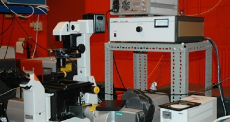
Multiphoton microscopy with fluorescence lifetime imaging
Multiphoton microscopy is combined with lifetime imaging to measure FRET in plant leaves, or measure the ultrafast dynamics of photosynthesis in leaves.
Mulitphoton microscopy (MP) combines confocal resolution with in depth imaging in turbid media. With the same system, second harmonic generation (SHG) from material with a noncentrosymmetric molecular structure can be detected (e.g. protein crystals). Typically, it is used to image multicellular systems like the roots or the leaves of plants, or strongly scattering media. As a special feature, it is equipped with fluorescence lifetime imaging (FLIM) detection.
Background
In the case that high photon densities are present, two or more low energy photons can be simultaneously absorbed in a single quantum event. Since the energy of a photon is inversely proportional to its wavelength, the two photons should be twice the wavelength necessary for single-photon excitation. It was until the beginning of the 1990s when Denk et al. introduced the two-photon laser scanning microscope resulting in the first biological applications of TPE. Because excitation in multiphoton microscopy occurs only at the focal point of a diffraction-limited spot, it is possible to create thin optical sections of thick biological specimens in order to obtain three-dimensional resolution.

Two-photon microscopy has some major advances over confocal microscopy for 3D imaging. First of all is the penetration of near-infrared light used for TPE much deeper than that of visible light used in conventional (SPE) confocal microscopy. TPE of thick biological samples allows imaging to a depth of more than 200 μm whereas SPE confocal microscopy is limited to depths of approximately 50 μm. In confocal microscopy fluorescent light is generated throughout the sample along the optical axis but only the signal from a thin focal plane is detected by placing an aperture (the so-called pinhole) at the image plane. However, by using TPE molecules are excited at the focal plane only and therefore no pinhole is required. In addition, TPE minimizes photobleaching and photodamage in out-of-focus regions that are usually limiting factors in conventional live cell imaging.
FLIM
Multiphoton microscopy is combined with fluorescence lifetime imaging (FLIM). FLIM monitors the distribution of the fluorescence lifetimes of a fluorophore at the different locations within the sample. The fluorescence of a sample is monitored as a function of time after excitation by a flash of light. We use the Time Correlated Single Photon Counting (TCSPC) method to record the fluorescence time trace for every pixel in the image. The lifetime may be sensitive to environmental factors like Ca2+ concentration, pH and polarity but is independent of dye concentration or light path length. The lifetime can be significantly reduced if excited state processes like FRET occur. Moreover, it can be used to measure the ultrafast dynamics of photosynthesis in leaves, algae or bacteria.
