
Nieuws
Possibilities of the electron microscopes of Wageningen Electron Microscope Centre (WEMC)
On 27 October Marcel Giesbers, Jan van Lent and Bart-Jan ten Hove presented the range of possibilities of the electron microscopes of Wageningen Electron Microscope Centre (WEMC), to a broad public of researchers gathered in Impulse at Wageningen University & Research (WUR). The facilities of WEMC are part of Wageningen Shared Research Facilities (CAT-AgroFood) and can be used by researchers of WUR, as well as by researchers from companies and other knowledge institutes.
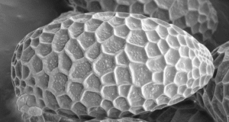
The electron microscopes can be used for studying multiple samples and products, such as those from biological origin, or those resulting from chemical of physical processes.
Marcel Giesbers started the seminar with an overview of the equipment for electron microscopy and sample preparation facilities, available at WEMC. A brief overview on the physical principles of electron microscopy, including the outline of the different characteristics and possibilities of SEM (scanning electron microscopy) and TEM (transmission electron microscopy) was presented. Also, information on related techniques and the various possibilities including the options for sample preparation was shown.

Finally, Marcel concluded with an overview of the different ways to use the facility , ranging from self-serve use, assisted use, full service use and combinations thereof.
WEMC provides training courses for those interested in self-serve use of the electron microscope facilities. Of course, WEMC also provides assistance for full service use of the WEMC facilities.
Traditionally, electron microscopy produces 2D images. The presentation of Jan van Lent focused on the techniques for 3D visualization at the nanoscale level, by using single particle cryo-electron microscopy or electron tomography. Electron tomography creates a series of tilted 2D images and subsequently recreates a 3D volume, by weighted back projection after alignment of the series. The procedures, requirements, data collection and the computerized reconstructions for electron tomography of a wide variety of samples were outlined and illustrated by with many beautiful photographs and movies.
Jan-Bart ten Hove ‘s presentation, with the intriguing title “Box-in-Box nanoassemblies: an investigation using cryoTEM” showed how nanoparticles are embedded in micelles.
The EM imaging clearly showed the fine details of both their morphology and structure these complex particles. The images are highly suited for both qualitative and quantitative analysis. Further information on the research of Jan-Bart ten Hove will be published soon.
The seminar was concluded with an informal discussion, where the guests had the opportunity to discuss their specific questions with the experts. The electron microscope facilities are located at the WEMC, the Wageningen Electron Microscope Centre.
If you are interested to know more about the microscopes of WEMC or other equipment of WUR, please see Shared Research Facilities for more information or or contact Shared Research Facilities directly.