
Equipment
Circular dichroism: steady-state and stopped-flow
Technique that is used to obtain structural information, for example about the arrangement of peptide bonds in secondary protein structure elements like helices and sheets.
Circular dichroism (CD) can be used to obtain structural information, for example about the arrangement of peptide bonds in secondary protein structure elements like helices and sheets. The amount of the different secondary structure elements can be obtained by fitting a CD spectrum. Instruments for normal and stopped-flow CD are available.
Principles Circular Dichroism (CD)
A molecule shows circular dichroism (CD) when it meets two requirements: the molecule should contain a chromophore and the molecule should be chiral. These molecules, for example proteins, interact differently with left- and right-handed circularly polarized light (see figure). The difference in absorbance of the left (EL)- and right-handed (ER) circularly polarized light is defined as: De = el – er. In this equation, el and er are the molecular extinction coefficients for the right and left circularly polarized beams of light. The CD spectrum of a protein molecule can be used to obtain structural information, for example about the arrangement of peptide bonds in secondary structure elements like helices and sheets. The amount of the different secondary structure elements can be obtained by fitting a CD spectrum to a set of known reference spectra. Some of these fit programs are available on the web (see Recommended literature and links).
Recommended literature and links:
- N.J. Greenfield, Analytical Biochemistry 235, 1 (1996)
- “Circular Dichroism – Principles and Applications” (K. Nakanishi, N. Berova and R.W. Woody, eds.), VCH, New York (1994)
- “Circular Dichroism and the Conformational Analysis of Biomolecules” (G.D. Fasman, ed.), Plenum Press, New York (1996)
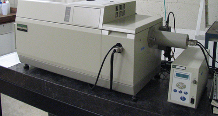
Jasco Spectropolarimeter J-715
High-precision instrument for CD measurements (manual). A powerful xenon lamp combined with a double monochromator enables accurate wavelength selection in the 165 to 900 nm range. The CD apparatus contains a photo elastic modulator (PEM) that generates circularly polarized light. Furthermore, it is possible to measure fluorescence detected CD. Cells of different sizes, ranging from a path length of 0.1 mm to 10 cm, can be used in the sample chamber. The temperature of the sample is controlled by a Peltier element (from 10 to 100 °C). Data recording and analysis is performed on a PC workstation, equipped with Jasco software (version 1.52.01).
Stopped-flow CD: Bio-Logic SFM-4
The Bio-Logic SFM-4 stopped flow apparatus contains four independently controlled syringes, which allows a minimal dead time of 0.7 milliseconds (manual). Four different optical modes can be used: absorbance, circular dichroism (CD), fluorescence and fluorescence anisotropy. The fluorescence anisotropy option uses the newly developed EMFA method (Excitation Modulated Fluorescence Anisotropy). This method requires neither mechanical polarizer rotation nor G-factor correction. Light is provided by a 150 W Xenon-Mercury lamp, and the wavelength is selected using a single monochromator. Two photomultipliers are available for simultaneous measurements of, for example, CD and fluorescence. Detection light is selected by emission filters (Andover). Data recording and analysis is performed on a PC workstation, equipped with Biokine software. Additional software is used to perform global analysis of experimental data.

Publications
- Georgakopoulou, S., Zwan, G. van der, Bassi, R., Grondelle, R. van, Amerongen, H. van & Croce, R. (2007). Understanding the changes in the circular dichroism of light harvesting complex IIupon varying its pigment composition and organization. Biochemistry, 46(16), 4745-4754.